Macular detachment in morning glory syndrome
Abstract
Purpose: The purpose of this study is to report a unique case of morning glory syndrome.
Methods: This study included ophthalmologic examination, optical coherence tomography and a review of the relevant literature.
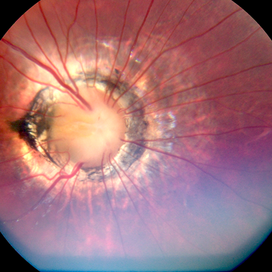
Result: A 7-year-old girl with a history of morning glory syndrome was periodically examined in our clinic for 5 years. Suddenly, she presented with the complaint of decreased vision. Examinations revealed macular detachment. The visual field of the affected eye was significantly narrowed. OCT also revealed the presence of a fibrous cord in the centre of the optic nerve, which protruded into the vitreous body.
Discussion: Morning glory syndrome is an uncommon congenital disorder characterized by a widely enlarged papilla that is pink-orange in colour, with a small glial tuft in the centre. The retinal vessels are arranged radially in relation to the papilla. A pigmented ring surrounds the excavation. The incidence is not well known. The effect is generally unilateral. This syndrome manifests as optic atrophy. However, the atrophy does not progress. Visual impairment sometimes occurs when macular detachment arises, as occurred in our patient. After 5 years of observation, our patient’s vision dramatically worsened as a result of macular detachment. There are various theories for the development of macular detachment in MGS: exudative, traction and rhegmatogenous8. No break was found in our patient, so the cause of the detachment was most likely the inflammatory process.
Copyright ©2021. All rights reserved.