Acute zonal occult outer retinopathy (AZOOR) progression: 30-year follow-up of a clinical case
Abstract
Purpose: The purpose of this study is to report a 30-year follow- up of a clinical case with progressive acute zonal occult outer retinopathy (AZOOR).
Methods: This study included an ophthalmic examination, optical coherence tomography (OCT), visual field studies, an electroretinogram (ERG) investigation, and a review of the relevant literature.
Results: A 34-year-old woman presented with a central scotoma in her right eye that had lasted for 5 months and a similar complaint in her left eye for a week. Changes in the fundus were minimal. The diagnosis was difficult. Later, as a result of the detection of a pronounced change in the ERG, local narrowing of the visual field (VF) and other symptoms, it was possible to make a diagnosis of AZOOR. We treated this patient first with parabulbar injections of dexamethasone and then with intramuscular ceftriaxone and intravenous dexamethasone. This treatment has had a stable positive effect.
Conclusion: AZOOR occurs suddenly, most often in women. Initially, minimal retinal changes are characteristic of this disease, accompanied by clearly defined areas of retinal dystrophy. There is a local narrowing of the visual field and a decrease in the amplitude of the ERG.
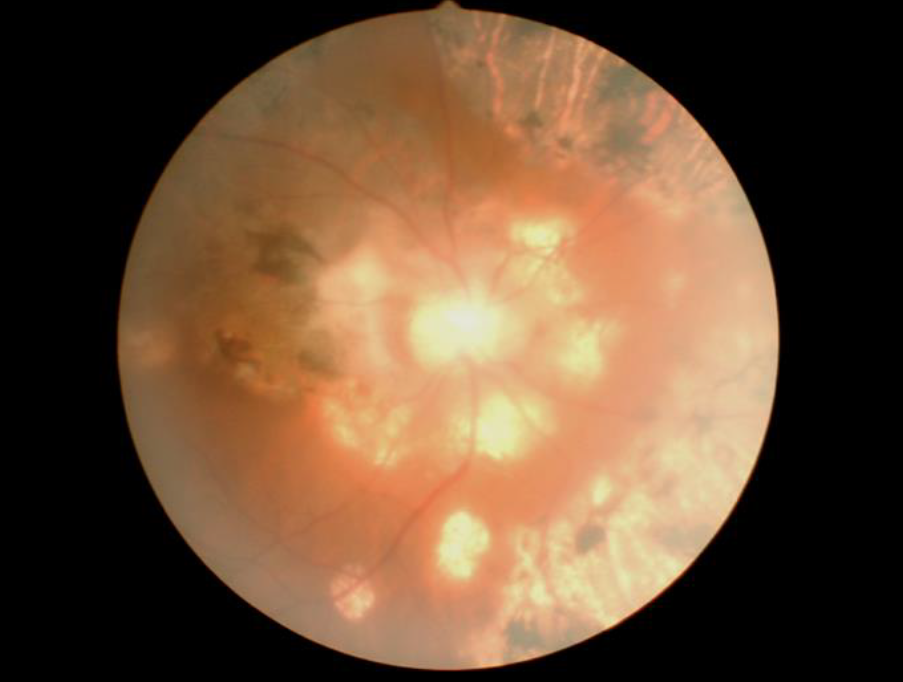
The difference between this disease and other diseases accompanied by a decrease in ERG is the good response to systemic corticosteroids (parabulbar or intravenous dexamethasone). The difference between this disease and other diseases accompanied by a decrease in ERG is the good response to systemic corticosteroids (parabulbar or intravenous dexamethasone).This disease is characterized by periodic exacerbations and gradual progression. After 30 years of observation of the present case, there was a significant decrease in visual acuity (VA) and the visual field (VF) and almost total retinal dystrophy, with the reduced ERG.


